Rarity Bioscience - Improved quality of life for patients
Technology
Ultra-sensitive detection of nucleic acid mutations for research and clinical use. Rarity offers ultrasensitive superRCA® assays for research, available for service testing. The superRCA® assays are designed for extreme sensitivity to enable the full potential of liquid biopsy.
Meny
Technology
Ultra-sensitivity – detects 1 mutation in 100,000 wild types
superRCA can detect 1 mutation out of a 100 000 wild-type DNA molecules.
The unprecedented sensitivity of superRCA is achieved by two consecutive Rolling Circle Amplification (RCA) reactions. A first standard RCA step is followed directly by a subsequent “in situ” Padlock Probing and RCA step. As a result the target region is genotyped with high specificity, enumerated with higher precision.
What is superRCA?
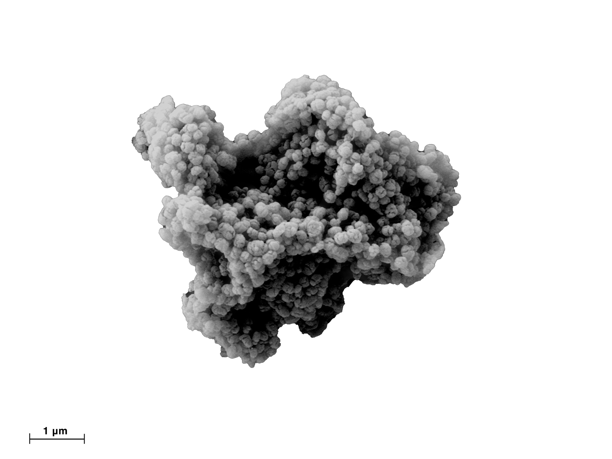
superRCA is an ultra-sensitive and highly specific molecular amplification technology. It is used to detect very small amounts of DNA sequence variants, like cancer mutations, in patient tissue and blood samples.
superRCA offers effective detection of multiple targets simultaneously – so called multiplexing. The assay can be performed in most hematology laboratories with existing equipment, using well-established flow cytometry for read-out, enabling more accessible testing with shorter response times.
The convenient, non-intrusive liquid biopsy-format enables higher frequency of testing for improved monitoring of molecular relapse and prevention of clinical relapse.
The same superRCA assays can be used to analyze both cell free and genomic DNA, making it suitable for a variety of liquid biopsy applications, such as bone marrow or blood samples for monitoring of leukemias, or using cfDNA derived from the plasma for monitoring of solid tumor cancers.
The assays can be used by clinicians and laboratories for tumor informed monitoring of patients. The workflow enables automation and uses only standard lab equipment.
For researchers and pharmaceutical companies, our services and research kits offer a convenient and efficient toolbox for investigating numerous therapeutic areas to design companion diagnostics and advance personalized medicine.
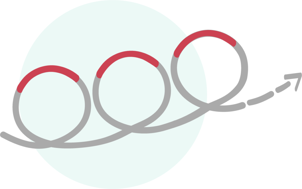
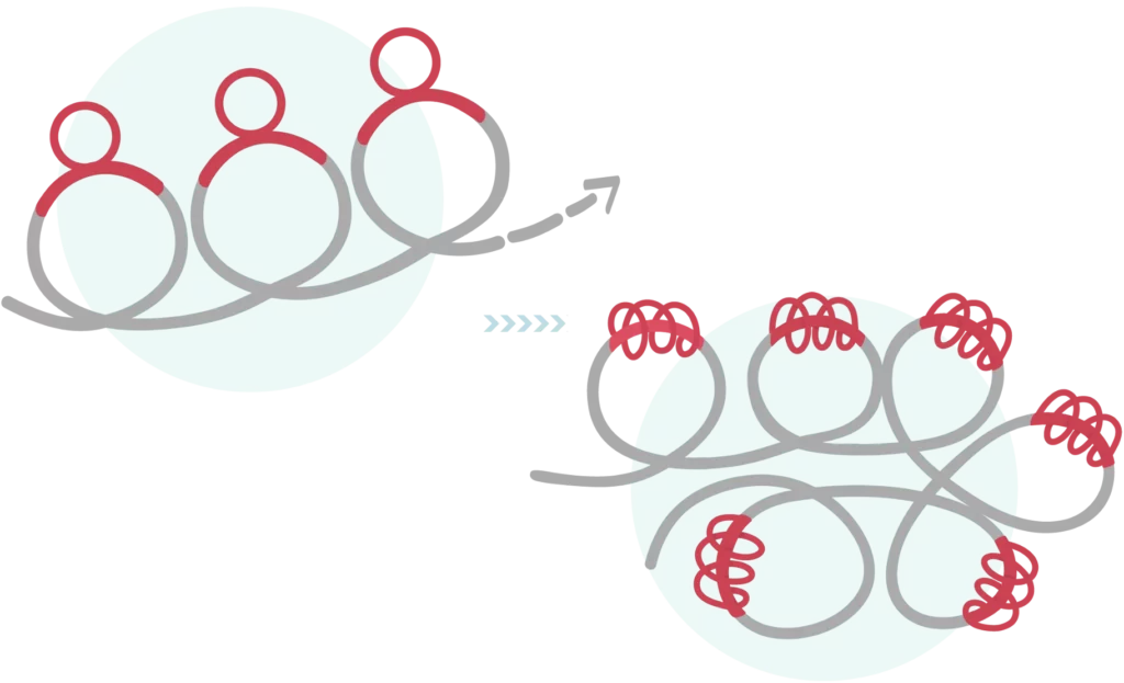
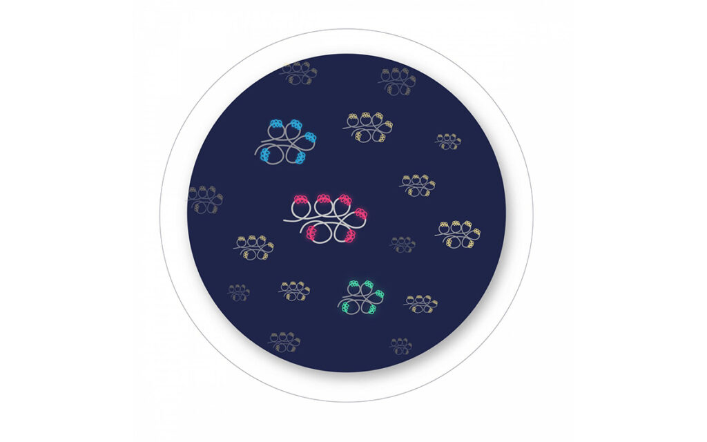
superRCA utilizes Rolling Circle Amplification (RCA) and Padlock probes in a novel way to achieve highly specific, ultra-sensitive detection of nucleic sequences. The method produces a relatively large, self-constrained structure – a superRCA structure – that can be directly analyzed by microscopy or automated using flow cytometry without the need for partitioning.
Flow cytometry with fluorescent labelling ensures that multiple targets can be analyzed simultaneously. As a result, the platform is extremely effective for patient-near, fast and cost-effective patient monitoring as well as companion diagnostics and pharmaceutical development .
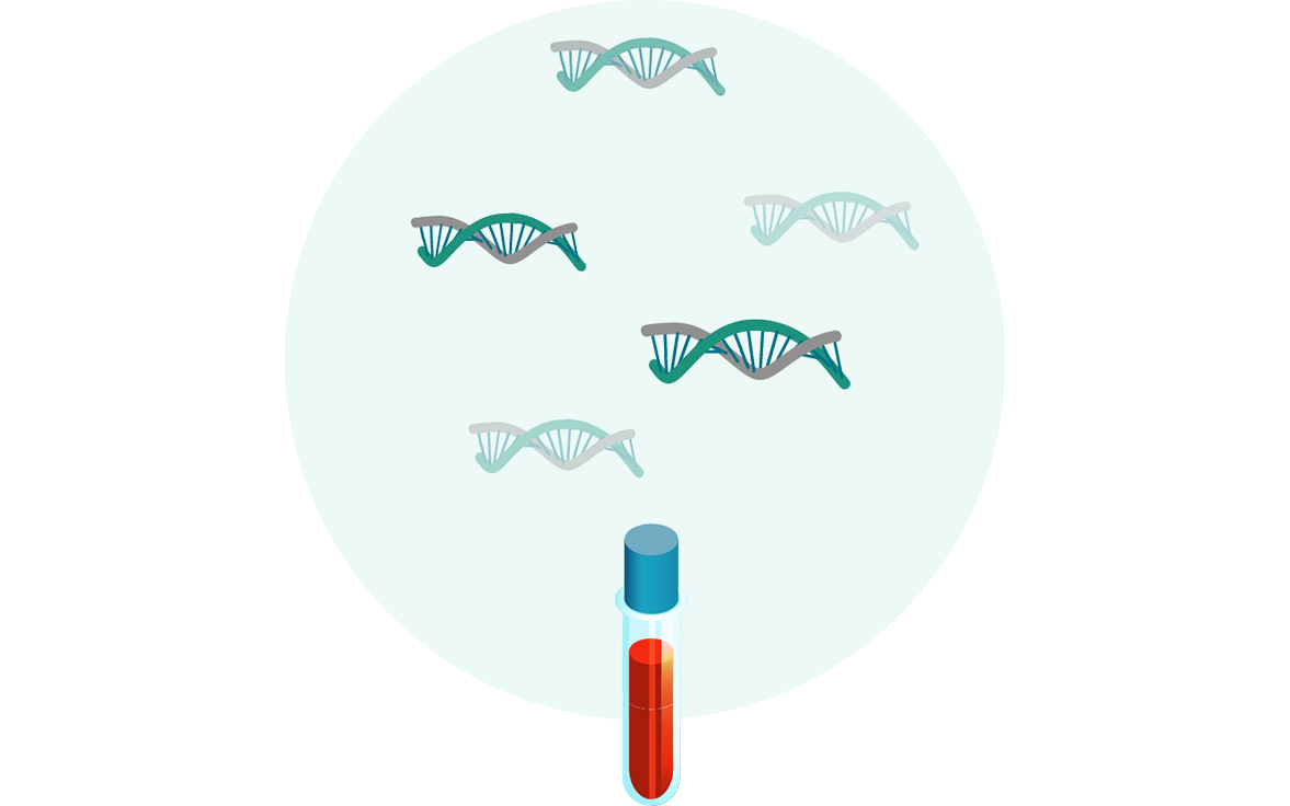
Step 1
DNA is extracted from the sample, either whole blood, bone marrow or tissue.
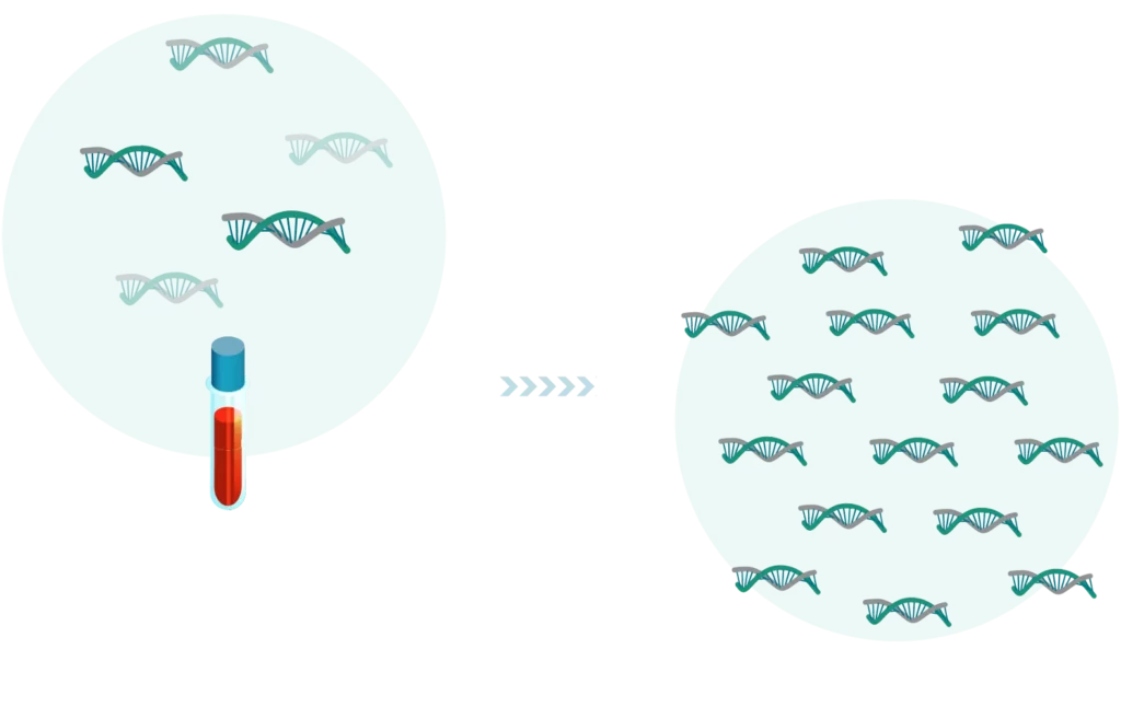
Step 2
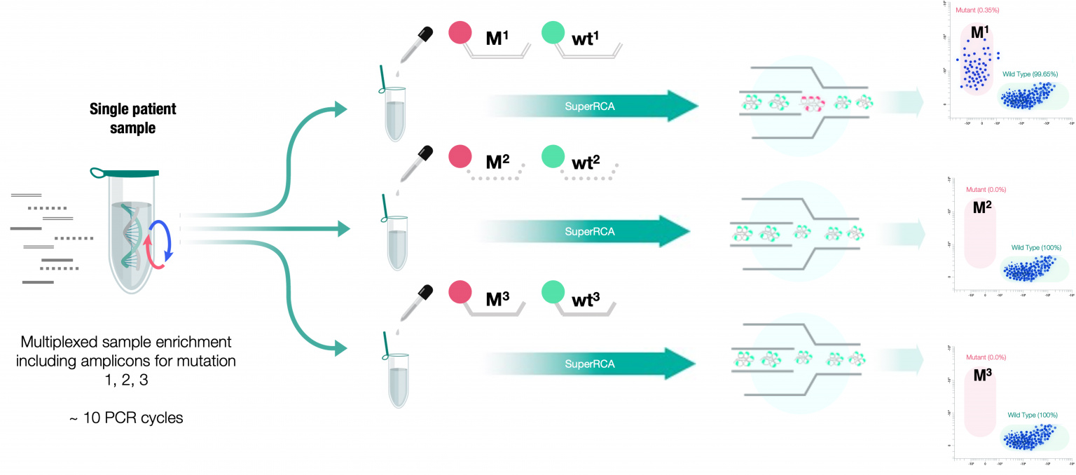
The DNA sequences of interest, known to be mutated in a patient’s malignant cells, are first enriched by a limited pre-PCR (~10 cycle) amplification.
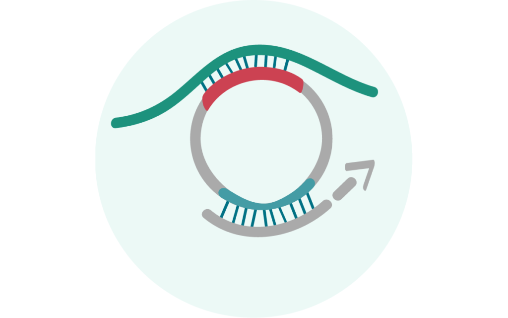
Step 3
The enriched sample then undergoes a ligase-mediated circularization of one strand.
Step 4
The circularized strands containing the target region are then amplified by the first Rolling Circle Amplification (RCA) step.
Step 5
This is followed by Padlock Probe ligation and a highly-specific, second RCA step. During this step, the second RCA encircles the first RCA product to form large superRCA structures that can be analyze by flow cytometry.
Step 6
The superRCA products can be scored as mutant- or wildtype-specific using fluorophore-labeled hybridization probes and recorded as individual, brightly fluorescent objects in a standard flow cytometer. For multiplex assays, analysis is similarly done by using multiple fluorophores and wavelengths.
Why it's different
Why superRCA makes a difference
Unprecedented ultra-sensitivity of 1 mutation in 100 000 wild types due to the novel combination of the extremely high specificity of Padlock probes and RCA
Meets or exceeds currently available technologies like ddPCR, NGS and traditional liquid biopsy assays by 10-100 times
Outstanding performance on High GC% targets. Assessment of high GC content with the same sensitivity as low GC content – with minimum bias.
High multiplexity creates opportunities for effective research, monitoring therapies and drug resistance, sensitive multiplex detection of mutations for treatment selection (companion diagnostics), quantitative measurement for patient follow up and clinical phase drug development
Convenient and reliable analysis by well-established flow cytometry for speed by patient-near testing.
Other read-out and analysis methods including fluorescence microscopy
Speed and cost-effectiveness at high-throughput.
An integrated sample-to-answer system with robotic automation using standard lab equipment and well-established instrumentation, minimizes manual handling and is highly amenable to clinical practice
Currently available as research use only kit or as a customized service
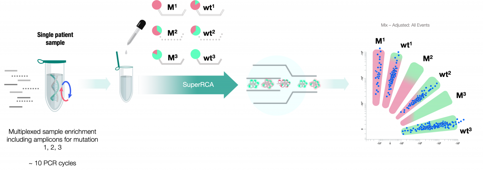
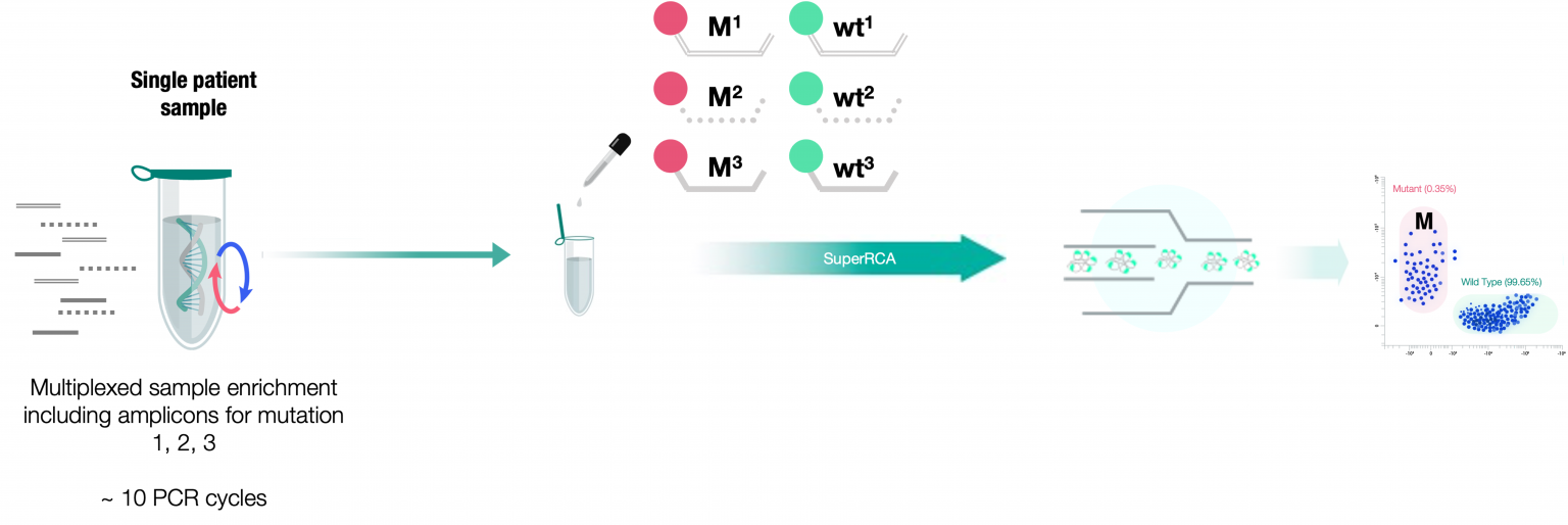
Multiplexing
The flexibility of multiplexing superRCA
The SuperRCA technology has intrinsic properties that allows for Multiplexing in a number of different ways based on application and user need. Multiplexing, in any form, means to be able to analyze a single sample for several sequences variations.
For all SuperRCA assays, the first step is an initial pre-amplification, were an assay specific amplicon is used for multiplexed target enrichment. This is undergoing approximately ten cycles of PCR, meaning enough to increase you initial sample to ensure statistical detection, but low enough to avoid PCR induced errors affecting the specificity. This step is comparable with library prep for sequencing.
Once the library prep is completed, the sample can undergo the SuperRCA incubation in a few different ways depending on the type of multiplex readout.
Parallel-Plex
For Parallel-Plex, the pre-amplified products are split into several wells and incubated with different padlock targets per well, meaning that each well is then analyzed for its individual target mutation. This can be done straight forward with only two colors for mutant and wildtype in each well.
Ratio-Labeling Multiplex
If high throughput and low running costs is important, we useRatio-Labeling Multiplex. Here the pre-amplified products remain in one single well, and different ratios of the two fluorophores are added to the different target probes. During the flow-cytometry read-out, presence of the different mutations will occur as cluster based on the specific ratios, and can be gated accordingly. This enables multiplex analysis while still only using two colors.
Multicolor Multiplex
For even higher throughput and cost efficiency, one can use MulticolorMultiplex, where different fluorophores are attached to each padlock probe, allowing simultaneous readout of multiple targets. This is limited to the number of lasers in the flow cytometer as well as the choice of dyes, and for additional expansion, multiple colors can be combined with ratio labeling to reach maximum multiplex.
Comboplex
The final option that can be combined with any of the above is Comboplex, where the same fluorophore is attached to multiple mutant probes, meaning that the readout will provide an answer if any of the mutations is present in the sample, but not particularly which one. This can be used for long term monitoring and/or in combination with a secondary drop-down assay in case sample is positive.
Rolling circle amplification (RCA) is an isothermal amplification method, which generates strands containing thousands of repeats complementary to the DNA circle that serve as the original template for the replication.
The single-strand clustered amplification products can be labeled with fluorophores or chromogenic functional groups by oligonucleotides hybridization. RCA can be used to to magnify detection events locally into highly visible signals, in combination with the molecular tools that generate circular reaction products like in situ PLA, padlock probes, selector probes, PLAYR etc.
A padlock probe is a short DNA oligonucleotide with segments at the 3’ and 5’ ends that are complementary to a target region. Upon hybridization, the two ends of the probe oriented in juxtaposition on the target template, leaving a nick site in the double-stranded structure. The nick site is sealed by a DNA ligase, and thereby the padlock probe is wound around and locked on the target strand. The DNA ligase activity is sensitive to base pair mismatches around the nick site, empowering the single base discrimination capacity of the padlock probes.
The central part of the padlock probe is not target complementary and can harbor specific sequences serving different purposes, such as a detection probe hybridization site, sites for amplification primer hybridization, and capture probe binding site. Padlock probes have been used in many applications, for example for copy number variation analysis (CNV), single nucleotide polymorphism (SNP) analysis, gene expression profiling, alternative splicing analysis and pathogen detection.
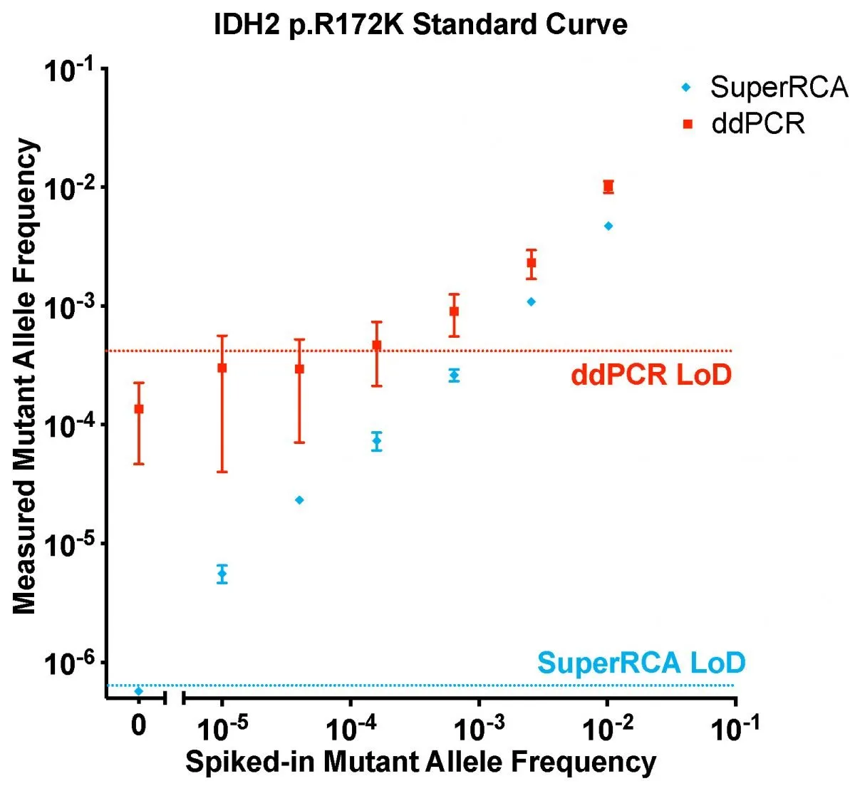
Figure 1. Spike in dilutaion curve comparing ddPCR with superRCA.
Taqman probe has been the fundamental element for the allele distinction function in various PCR based mutation detection technologies. In emulsion/microcompartment digital PCR (dPCR) and quantitative PCR (qPCR), the Taqman probe distinguishes different alleles during PCR amplification steps. However, the hydrolysis feature in the Taqman probe relies on the 5’ to 3’ exonuclease possessed by the PCR polymerases, thus limits the selection of PCR polymerase to Taq DNA poly which is suboptimal in PCR replication fidelity.
Other technologies for allele distinction such as BEAMing assays employs a bead-based amplification which allows to use high fidelity DNA polymerase to generate enough target molecules in bead and perform Taqman probe based allele distinction after the PCR amplification. But it is quite tricky to ensure (one input molecule + bead) per emulsion to generate single molecule derived bead colony resulting excessive number of empty beads during enumeration.
All Taqman probe-based allele distinction relies on the hybridization affinity difference between the match/unmatch target so that only the matched probe give rise to fluorescent signals while the unmatched will fall off. However, the Gibbs free energy difference (ΔG) attributed by the single nucleotide (SNP distinction) heavily influenced by the environmental sequences (the sequence that Taqman recognizes), which determines the severity of missing binding of the Taqman probe.
In superRCA technology-based mutation detection assays, we use padlock probe based allele distinction mechanism. The Padlock probe utilize four interdependent mechanism to improve the genotyping sensitivity: 1. Probe hybridization (binding to the matched target), 2. Ligase mediated allele distinction (only perfect match base pair can be ligated into replicable circles). 3. Circle confirmation (only successfully ligated probe can contribute to a 2nd rolling and yield fluorescent signal while the miss matched probe contributes nothing to the total fluorescence), 4. Major voting mechanism (the concatemer based padlock probing employs hundreds to thousands of padlock probe to sense the same target molecule).
These four mechanisms enable extreme high fidelity allele distinction that is used in the superRCA based assays to provide cutting edge sensitivity.
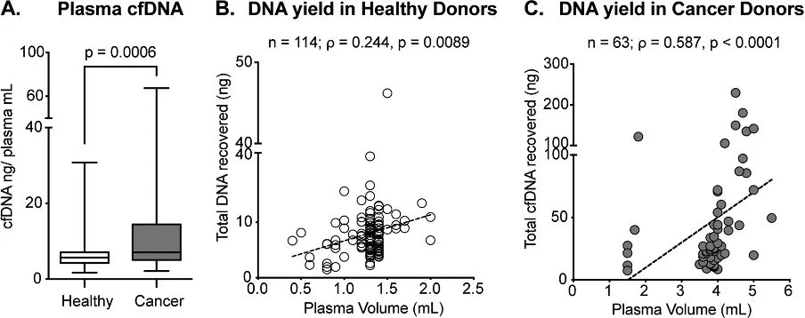
The total DNA recovered from different tissues varies depending on type of sample, source, and health status of the patient. Solid biopsies from mammalian tissues typically yield 0.2-0.4 mg genomic DNA (gDNA)/mg tissue. The amount depends on how many nucleated cells the tissue contains per mg and fatty tissues such as brain, bone marrow or tissues with high levels of extracellular matrix such as connective tissues typically fall in the lower range.
Blood, a connective tissue with a dilute, liquid extracellular matrix, produces the lowest gDNA yields – typically between 15-50 ng gDNA/mg (or 15-50 mg gDNA/ml) with an assumed average of 35, corresponding to around 5 million nucleated white blood-cells/ml in healthy individualA normal 10ml venous draw gives around 350mg gDNA, equivalent to around 100million haploid cells.
Even though blood is dilute with respect to gDNA, it is a readily available, non-invasive biological sample that is easily extracted from patients. Except for gDNA derived from nucleated cells, blood also contain circulating cell-free DNA (cfDNA) from recycled cells in the body. This pool of small, double stranded extracellular DNA fragments potentially reflects the genotypes of all the cells in the body and the concentration typically varies between 0-100 ng cfDNA/mlwith an average around 30 ng/ml for cancer patients. cfDNA is unstable with a reported half-life between 16 min to 2.5 h in circulation.
A normal 10ml venous drawgives around 150ng cfNDA, equivalent to around 45,500 haploid cells.
This means that in order to reliable sensitivity of 1:100,000, one would need 200,000 copies and hence 100,000 diplod cells, being 660ng cfDNA and hence around 40ml whole blood.
References:
Elazezy M, Joosse SA. Techniques of using circulating tumor DNA as a liquid biopsy component in cancer management. Comput Struct Biotechnol J. 2018 Oct 9;16:370-378. doi: 10.1016/j.csbj.2018.10.002. PMID: 30364656; PMCID: PMC6197739.
Anatoli Kustanovich, Ruth Schwartz, Tamar Peretz & Albert Grinshpun (2019) Life and death of circulating cell-free DNA, Cancer Biology & Therapy, 20:8, 1057-1067, doi: 10.1080/15384047.2019.1598759
Alborelli, I., Generali, D., Jermann, P. et al. Cell-free DNA analysis in healthy individuals by next-generation sequencing: a proof of concept and technical validation study. Cell Death Dis 10, 534 (2019). https://doi.org/10.1038/s41419-019-1770-3
This website uses cookies so that we can provide you with the best user experience possible. Cookie information is stored in your browser and performs functions such as recognising you when you return to our website and helping our team to understand which sections of the website you find most interesting and useful.
Strictly Necessary Cookies
Strictly Necessary Cookie should be enabled at all times so that we can save your preferences for cookie settings.
If you disable this cookie, we will not be able to save your preferences. This means that every time you visit this website you will need to enable or disable cookies again.